All published articles of this journal are available on ScienceDirect.

Perioperative Anesthetic Management of Wilms Tumor in a Child with Fanconi Anemia and Horseshoe Kidney: A Case Report

Abstract
Background
Fanconi anemia (FA) is a rare inherited bone marrow failure syndrome with multisystem congenital anomalies and a markedly elevated risk of malignancy. Children with FA undergoing major oncological surgery present significant anesthetic challenges, including severe pancytopenia, structural airway anomalies, congenital cardiac defects, and amplified hemorrhagic diathesis.
Case Presentation
We report the perioperative management of a 4-year-old, 12 kg boy with FA (FANCD1/BRCA2 biallelic mutation), a 7×5 cm Wilms tumor (Stage III) in the right moiety and isthmus of a horseshoe kidney, a Type IV membranous ventricular septal defect (VSD), and spina bifida occulta. After six cycles of neoadjuvant chemotherapy (vincristine-actinomycin-D), the child underwent radical tumor resection with isthmectomy under general anesthesia. Preoperative hematological optimization included packed red blood cell (PRBC) and platelet transfusion. The patient underwent videolaryngoscopy-facilitated intubation due to micrognathia. Invasive hemodynamic monitoring, Tranexamic acid infusion, and blood component therapy were employed to mitigate hemorrhagic risk. Ultrasound-guided bilateral posterior quadratus lumborum (QL) blocks (0.3 mL/kg of 0.25% ropivacaine per side) provided effective opioid-sparing analgesia, with Face, Legs, Activity, Cry, Consolability (FLACC) scores ≤3 for 18–20 hours, avoiding non-steroidal anti-inflammatory drugs (NSAIDs) given the potential chemotherapy-related nephrotoxicity risk. Estimated blood loss was 400 mL (~30% blood volume), managed with 200 mL PRBC and one platelet unit. The child was extubated awake in the operating theatre and discharged on postoperative day 10 with intact renal function.
Conclusion
Meticulous multidisciplinary preoperative planning, video-assisted airway management, goal-directed hemostatic therapy, and ultrasound-guided fascial plane analgesia were integral to safe perioperative management in pediatric FA undergoing high-risk oncological surgery.
1. INTRODUCTION
Fanconi anemia (FA) is the most common inherited cause of aplastic anemia in children, with an estimated incidence of 1–5 per million live births [1]. It results from biallelic mutations in at least 22 FANC genes that encode the FA-BRCA DNA damage repair pathway. The FANCD1 subtype, caused by biallelic BRCA2 mutations, is associated with a severe phenotype and a very high lifetime risk of hematological malignancies and solid tumors, including Wilms tumor [2, 3].
Anaesthetic management in FA patients is complicated by severe thrombocytopenia and anemia, craniofacial dysmorphism predisposing to a difficult airway, congenital cardiac defects, and immunosuppression from bone marrow failure [4]. Wilms tumor in a horseshoe kidney further amplifies intraoperative hemorrhagic risk owing to complex aberrant multi-vessel arterial supply arising from the aorta and iliac vessels [5]. Ultrasound-guided regional anesthesia (UGRA) offers opioid-sparing analgesia with a favorable safety profile [6] and represents an especially important strategy where NSAIDs and high-dose systemic opioids are contraindicated, in FA patients where nephrotoxicity from prior chemotherapy may compromise long-term renal reserve. Reports of UGRA specifically within this context remain scarce. We present this case to highlight a structured multimodal approach with the posterior quadratus lumborum (QL) block as the primary analgesic modality.
2. CASE REPORT
The patient's parents provided written informed consent for the publication of this case report, including all clinical data, investigations, and intraoperative images, before submission. A 4-year-old boy weighing 12 kg presented for resection of a 7×5 cm Wilms tumor arising from the right moiety of a horseshoe kidney and extending to the isthmus and lower pole of the left kidney (Stage III), confirmed on 3D magnetic resonance imaging (MRI). Genetic testing identified a FANCD1/BRCA2 biallelic mutation (Fig. 1). Relevant comorbidities included a hemodynamically insignificant Type IV membranous VSD (left-to-right shunt; no pulmonary hypertension on echocardiography) and lumbosacral spina bifida occulta. Six cycles of neoadjuvant chemotherapy (vincristine and actinomycin-D; International Society of Paediatric Oncology [SIOP]-2001 protocol) had been completed before surgery. He also had a history of a previous surgery for hip dysplasia 3 years back under general anesthesia during which a Cormack-Lehane Grade IIIb direct laryngoscopy view was noted.
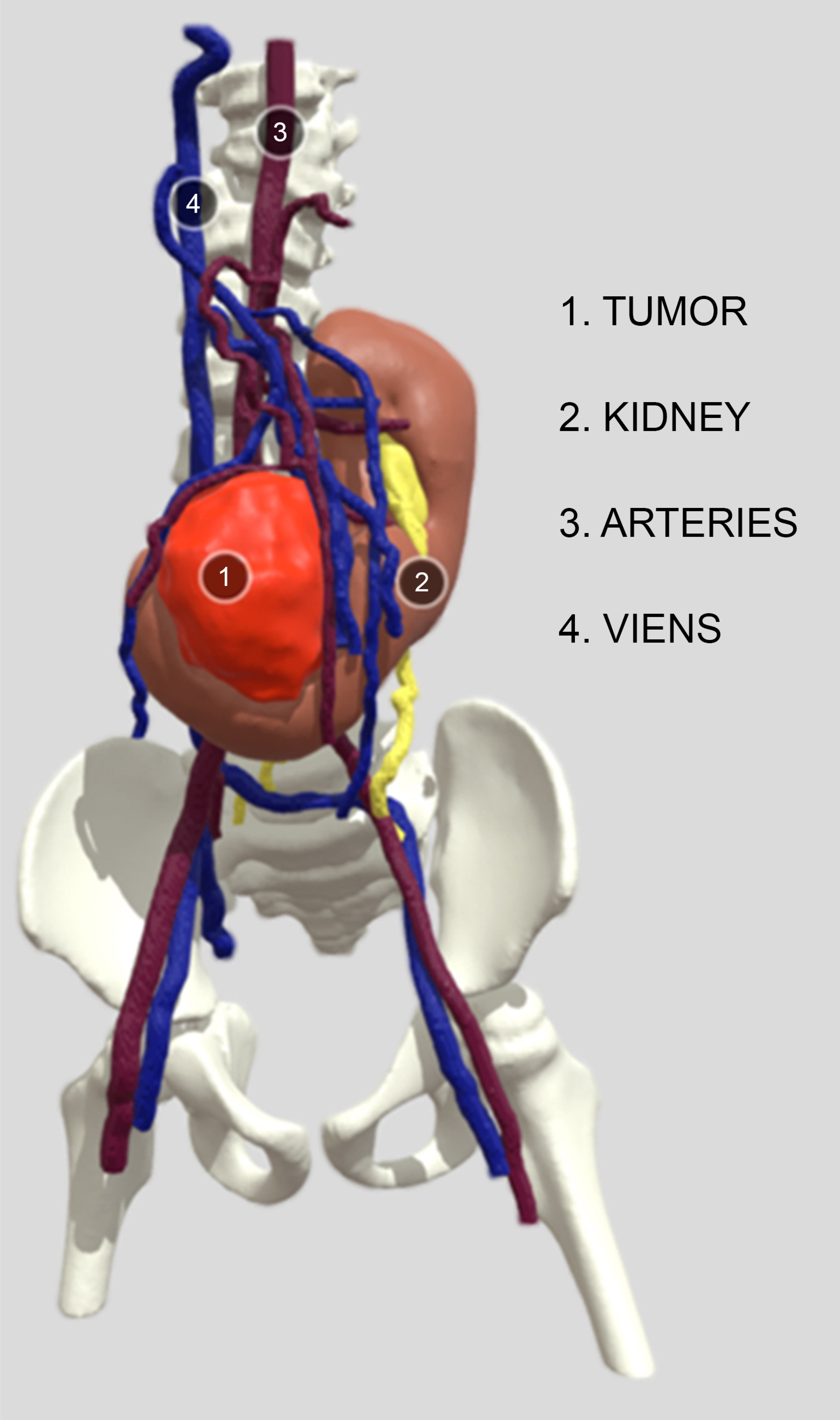
Three-dimensional magnetic resonance imaging reconstruction demonstrating the complex vascular anatomy of the horseshoe kidney. The Wilms tumor (1, red) occupies the right renal moiety and isthmus, with the remnant horseshoe kidney parenchyma (2, brown) visible medially. Aberrant arterial supply (3, red vessels) arises from the aortic bifurcation, with venous drainage (4, blue vessels) coursing bilaterally into the iliac system, illustrating the surgical hemorrhagic risk inherent to this anatomical variant.
Preoperative blood work revealed hemoglobin 6.8 g/dL, platelets 42,000/µL, and white cell count 2,100/µL, and reticulocyte count 0.4% (absolute reticulocyte count 16,000/µL, consistent with hypoproliferative bone marrow failure); serum creatinine was 0.5 mg/dL. Physical examination did not reveal petechiae or active mucosal bleeding at the time of preoperative assessment, although the platelet count of 42,000/µL represented a significant hemorrhagic risk for major surgery. Airway assessment demonstrated micrognathia, inter-incisor distance 2.5 cm, and Mallampati Class III. MRI confirmed anomalous vasculature arising from the aortic bifurcation and bilateral iliac vessels (Table 1). American Society of Anesthesiologists (ASA) physical status was III. Over 48 hours preoperatively, packed red cell transfusion (10 mL/kg) raised hemoglobin to 9.2 g/dL, and platelet transfusion raised the count to 78,000/µL. Prophylactic antibiotics (co-amoxiclav) and antifungal therapy (fluconazole) were prescribed given the neutropenia. Written informed consent for anesthesia and surgery was obtained from both parents/legal guardians before the procedure.
| Investigation | Result | Reference Range | Interpretation |
|---|---|---|---|
| Haematology | |||
| Haemoglobin (g/dL) | 6.8 | 11.5 – 13.5 | Very Low |
| Haemoglobin post-transfusion (g/dL) | 9.2 | 11.5 – 13.5 | Low |
| Hemoglobin postoperative Day 1 (g/dL) | 9.8 | 11.5 – 13.5 | Low |
| Total WBC count (/µL) | 2,100 | 4,000 – 11,000 | Very Low |
| Absolute Neutrophil Count (/µL) | 800 | 1,800 – 7,700 | Very Low |
| Platelet count (/µL) | 42,000 | 1,50,000 – 4,00,000 | Very Low |
| Platelet count post-transfusion (/µL) | 78,000 | 1,50,000 – 4,00,000 | Low |
| Hemoglobin postoperative Day 1 (g/dL) | 9.8 | 11.5 – 13.5 | Low |
| MCV (fL) | 102 | 75 – 87 | High |
| Coagulation | |||
| Prothrombin Time / PT (sec) | 13.2 | 11 – 14 | Normal |
| INR | 1.1 | 0.9 – 1.1 | Normal |
| aPTT (sec) | 34 | 28 – 40 | Normal |
| Renal Function Tests | |||
| Blood Urea Nitrogen / BUN (mg/dL) | 14 | 7 – 18 | Normal |
| Serum Creatinine (mg/dL) | 0.5 | 0.3 – 0.7 | Normal |
| Serum Creatinine postoperative Day 7 (mg/dL) | 0.5 | 0.3 – 0.7 | Normal |
| Serum Sodium (mEq/L) | 136 | 135 – 145 | Normal |
| Serum Potassium (mEq/L) | 3.9 | 3.5 – 5.0 | Normal |
| Liver Function Tests | |||
| SGOT / AST (U/L) | 38 | < 40 | Normal |
| SGPT / ALT (U/L) | 34 | < 40 | Normal |
| Serum Albumin (g/dL) | 3.4 | 3.5 – 5.0 | Low |
| Total Bilirubin (mg/dL) | 0.6 | 0.1 – 1.0 | Normal |
| Cardiac Investigations | |||
| ECG | Sinus rhythm; mild right-axis deviation | — | Abnormal |
| Echocardiography | Type IV membranous VSD (5 mm, L→R); no PH; LVEF 62% | — | Abnormal |
| Chest X-Ray | Mild cardiomegaly (CTR 0.52); no pulmonary oedema | — | Abnormal |
| Imaging & Special Investigations | |||
| Abdominal MRI | 7×5 cm Wilms tumour right moiety horseshoe kidney; aberrant multi-vessel supply from aortic bifurcation and bilateral iliac vessels | — | Abnormal |
| Renal DTPA Scan | GFR left kidney 42 mL/min; right kidney 26 mL/min (total GFR 68 mL/min/1.73 m2) | — | Abnormal |
| Genetic Testing (FANC panel) | FANCD1/BRCA2 biallelic pathogenic mutation confirmed | — | Abnormal |
Abbreviations: ALT = alanine aminotransferase; ANC = absolute neutrophil count; aPTT = activated partial thromboplastin time; AST = aspartate aminotransferase; BUN = blood urea nitrogen; CTR = cardiothoracic ratio; DTPA = diethylenetriaminepentaacetic acid; ECG = electrocardiogram; GFR = glomerular filtration rate; INR = international normalised ratio; L→R = left-to-right shunt; LVEF = left ventricular ejection fraction; MCV = mean corpuscular volume; MRI = magnetic resonance imaging; PH = pulmonary hypertension; PT = prothrombin time; VSD = ventricular septal defect; WBC = white blood cell count.
Preoperative vitals on the day of surgery were: heart rate 102 beats/min, blood pressure 88/54 mmHg (mean arterial pressure [MAP] 65 mmHg), respiratory rate 24 breaths/min, SpO2 97% on room air, and temperature 36.6°C; height was 98 cm and weight 12 kg. After oral midazolam premedication (0.3 mg/kg), standard monitoring (electrocardiography, pulse oximetry, non-invasive blood pressure, and capnography) was applied. Inhalational induction was performed with Sevoflurane (8% end-tidal titrated to loss of consciousness) in 100% oxygen. Rocuronium (0.6 mg/kg) was administered, and videolaryngoscopy (C-MAC® D-Blade, Karl Storz) was performed, converting a Cormack-Lehane Grade IIIb direct laryngoscopy view to Grade I; a 4.5 mm cuffed endotracheal tube was placed on the first attempt. Right radial arterial line (22G) and right internal jugular central venous catheter (4Fr double-lumen) were placed under real-time ultrasound guidance. Anesthesia was maintained with Sevoflurane (0.8–1.2 minimum alveolar concentration [MAC]) in an oxygen–air mixture (fraction of inspired oxygen [FiO2] 0.5), fentanyl infusion (1–2 µg/kg/h), and intermittent rocuronium boluses. Normothermia was maintained throughout with a forced-air warming blanket and warmed intravenous fluids. Post-induction baseline hemodynamics were: heart rate 98 beats/min, invasive BP 82/48 mmHg (MAP 59 mmHg), central venous pressure (CVP) 7 cmH2O, and SpO2 99%. Baseline arterial blood gas (ABG) at this point showed: pH 7.38, PaO2 198 mmHg (FiO2 0.5), PaCO2 38 mmHg, HCO3− 22 mEq/L, lactate 1.1 mmol/L, hematocrit 28%, ionized calcium 1.12 mmol/L, and blood glucose 86 mg/dL.
Prior to surgical incision, with the child supine, bilateral posterior QL blocks were performed under ultrasound guidance using a low-frequency curvilinear transducer (2–5 MHz). The quadratus lumborum muscle was visualized in a transverse oblique view. A 22G, 50 mm echogenic needle was advanced in-plane to deposit 0.3 mL/kg of 0.25% ropivacaine posterior to the QL muscle, targeting the thoracolumbar fascia; real-time craniocaudal spread within the fascial plane was confirmed sonographically. Blocks were completed bilaterally, with total ropivacaine dose within safe pediatric limits (≤2 mg/kg). NSAIDs were withheld throughout. Tranexamic acid was administered as a loading dose of 15 mg/kg over 10 minutes, followed by an intraoperative infusion of 2 mg/kg/h.
Surgery lasted six hours and comprised radical tumor resection with isthmectomy and regional lymph node sampling. The highest-risk phase, mobilization of the aberrant tumor vasculature at approximately the third hour, was marked by a transient rise in heart rate to 138 beats/min and a dip in MAP to 52 mmHg, managed with a bolus of crystalloid (10 mL/kg) and a brief noradrenaline infusion (0.05 µg/kg/min) titrated off within 20 minutes. CVP ranged between 6 and 10 cmH2O throughout, rising to 11 cmH2O following transfusion. Mid-surgical ABG (3 hours) showed: pH 7.34, PaCO2 40 mmHg, lactate 1.8 mmol/L, hematocrit 21%, ionized calcium 1.04 mmol/L (corrected with 10% calcium gluconate 0.2 mL/kg), and glucose 92 mg/dL. Estimated blood loss was 400 mL (approximately 30% of estimated blood volume for a 12 kg child); 200 mL PRBC and one unit of platelets were transfused. Pre-closure ABG showed: pH 7.37, PaCO2 38 mmHg, lactate 1.3 mmol/L, and hematocrit 26% following transfusion. Mean urine output was 1.8 mL/kg/h. Core temperature was maintained at 36.2–36.8°C. Pre-reversal train-of-four (TOF) count was 2/4; sugammadex (2 mg/kg) was administered, and TOF ratio confirmed ≥0.9 before extubation. The child was extubated awake in the operating theatre with SpO2 99% on 4 L/min supplemental oxygen and shifted to the pediatric intensive care unit (ICU) for postoperative monitoring.
Postoperatively, bilateral QL blocks provided effective analgesia assessed at defined time points: FLACC scores were 2 at 1 hour, 2 at 6 hours, 3 at 12 hours, 3 at 18 hours, and 4 at 24 hours, at which point rescue analgesia was escalated. Total fentanyl consumption in the first 24 hours was 0.5 mg/kg, reflecting a clinically meaningful opioid-sparing effect. Rescue analgesia comprised intravenous paracetamol (15 mg/kg 6-hourly) and fentanyl boluses (0.5 µg/kg as needed). General postoperative care included continuation of prophylactic antibiotics (co-amoxiclav) and antifungal therapy (fluconazole) until neutrophil recovery, daily complete blood count monitoring, strict fluid balance with hourly urine output monitoring targeting ≥1 mL/kg/h, granulocyte colony-stimulating factor (G-CSF) was withheld per oncology guidance given FA-related bone marrow fragility, thromboprophylaxis was deferred given the thrombocytopenia, and oncology follow-up was arranged for resumption of adjuvant therapy planning. Postoperative SpO2 remained ≥97% on room air from hour 4 onwards; respiratory rate normalized to 22–24 breaths/min. No block-related adverse events were observed. Postoperative day 1 blood work showed hemoglobin 9.8 g/dL and platelets 64,000/µL. Creatinine trend was: 0.5 mg/dL (baseline), 0.6 mg/dL (day 1), 0.5 mg/dL (day 3), 0.5 mg/dL (day 7), and 0.5 mg/dL (discharge day 10), confirming intact renal function throughout. The child was transferred from the pediatric ICU to the ward on postoperative day 1 and discharged on day 10 with normal urine output and no complications.
3. DISCUSSION
This case highlights the convergence of hematological, airway, surgical, and analgesic challenges inherent to pediatric FA patients undergoing major oncological surgery, and demonstrates that a structured multidisciplinary approach yields favorable outcomes.
Preoperative hematological optimization is the foundation of safe surgical management in FA. Progressive bone marrow failure results not only in quantitative but also in qualitative platelet defects, making transfusion thresholds alone insufficient guides [4, 7]. We targeted a hemoglobin >9 g/dL and platelets >75,000/µL before surgery, consistent with established guidelines for major pediatric surgery [8]. Intraoperative Tranexamic acid reduced antifibrinolytic activity and the risk of bleeding [9].
Craniofacial anomalies, including micrognathia, occur in up to 50% of FA patients and predict difficult laryngoscopy [4]. Videolaryngoscopy is recommended as the first-line technique for anticipated difficult pediatric airways, converting a Cormack-Lehane Grade III direct laryngoscopy view to Grade I without repeated airway instrumentation and the risk of mucosal hemorrhage [10]. Ultrasound-guided arterial and central venous placement minimized needle passes and, therefore, infectious and hemorrhagic complications, which are critically important in a neutropenic and thrombocytopenic child [6].
The posterior QL block was selected as the primary analgesic modality for several reasons. First, due to the presence of spina bifida occulta, caudal block was avoided, and QL block is superior to caudal blocks [11, 12]. Second, unlike transversus abdominis plane (TAP) blocks, the posterior QL approach targets the thoracolumbar fascia, with cadaveric studies demonstrating spread towards the paravertebral space, thereby providing both somatic analgesia for the flank incision and potential visceral coverage relevant to retroperitoneal dissection [13]. This dual coverage is important in Wilms tumor surgery, where both incisional and renal bed nociception contribute significantly to the pain burden. Published pediatric experience confirms that posterior QL blocks at 0.25–0.3 mL/kg of ropivacaine provide 16–24 hours of effective analgesia in abdominal surgery, consistent with our observed FLACC scores [14]. Third, NSAIDs were absolutely contraindicated given the risk of nephrotoxicity from prior vincristine and actinomycin-D chemotherapy, and the hemorrhagic diathesis of FA; renal function was normal at baseline (creatinine 0.5 mg/dL, GFR 68 mL/min/1.73 m2), but preservation of renal reserve was a priority; the QL block offered a safe and effective alternative [6].
Hypothermia coagulopathy, which is catastrophic in a thrombocytopenic patient, was prevented by active warming throughout the six-hour procedure [15]. Anaesthetic goals for the coexisting VSD focused on preventing pulmonary hypertension by maintaining normoxia, normocarbia, and normothermia to avoid shunt reversal [13]. Strict aseptic technique for all invasive procedures and ultrasound guidance reduced the number of needle passes and infectious risk in the context of neutropenia [6].
This report carries the inherent limitations of a single case description. First, thromboelastography (TEG)- guided transfusion could have been used in this patient to avoid unnecessary component use and its immunomodulatory sequelae in an already immunocompromised host. However, it was not used due to its unavailability in our institution. Second, the FANCD1/BRCA2 subtype represents one of the rarer and more severe FA genotypes, and the anaesthetic challenges encountered may not be fully generalizable to children with FA caused by mutations in other FANC genes, who may have less severe hematological or structural phenotypes. Third, the duration of QL block analgesia was inferred from the FLACC score trajectory and fentanyl consumption rather than confirmed by formal sensory assessment, which is inherently difficult to perform in a 4-year-old child postoperatively. Fourth, long-term oncological and renal functional outcomes beyond the index hospital admission are not available for this report.
CONCLUSION
Safe anaesthetic management of pediatric FA for major oncological surgery demands aggressive hematological optimization, videolaryngoscopy-assisted airway management, goal-directed hemostatic therapy, and ultrasound-guided regional analgesia. The posterior quadratus lumborum block is a valuable opioid-sparing tool offering somatic and visceral analgesic coverage in retroperitoneal surgery, particularly when NSAIDs and systemic opioid infusions carry unacceptable risk. Multidisciplinary tumor board planning and institution-specific FA protocols are essential to optimize outcomes in this rare, high-risk population.
AUTHORS’ CONTRIBUTIONS
The authors confirm their contributions to the paper as follows: S.M.: Conceptualization, Anaesthetic Management, Writing – Original Draft, Writing – Review & Editing; K.S.S.: Anaesthetic Management, Writing – Original Draft. KBTK: Anaesthetic Management, Data Collection, Writing – Review & Editing. All the authors have read and approved the final manuscript.
LIST OF ABBREVIATIONS
| ABG | = Arterial Blood Gas |
| ASA | = American Society of Anesthesiologists |
| BMI | = Body Mass Index |
| BP | = Blood Pressure |
| CVP | = Central Venous Pressure |
| ETCO₂ | = End-Tidal Carbon Dioxide |
| FA | = Fanconi Anemia |
| FLACC | = Face Legs Activity Cry Consolability |
| FiO₂ | = Fraction of Inspired Oxygen |
| HR | = Heart Rate |
| MA | = Maximum Amplitude |
| MAC | = Minimum Alveolar Concentration |
| MAP | = Mean Arterial Pressure |
| MRI | = Magnetic Resonance Imaging |
| NSAIDs | = Non-Steroidal Anti-Inflammatory Drugs |
| PRBC | = Packed Red Blood Cells |
| QL | = Quadratus Lumborum |
| SpO₂ | = Peripheral Oxygen Saturation |
| TEG | = Thromboelastography |
| TOF | = Train-of-Four |
| UGRA | = Ultrasound-Guided Regional Anesthesia |
| VSD | = Ventricular Septal Defect |
CONSENT FOR PUBLICATION
Written informed consent for publication of this case report, including all clinical details and associated data, was obtained from the patient's parents/legal guardians. The original consent forms are retained by the corresponding author and will be provided to the editorial office upon request.
AVAILABILITY OF DATA AND MATERIALS
All data relevant to this case report are included within the article. The datasets used and/or analyzed are available from the corresponding author [S.M.] on reasonable request.
ACKNOWLEDGEMENTS
The authors thank the pediatric surgery and the pediatric ICU teams at KIMS, India for their collaboration, and the patient's family for their trust and consent.